As most people realise, the eye examination should be a part of every persons’ regular health checks.
Apart from sight correction, regular examinations allow our optometrists to inspect your eyes for
numerous conditions that affect general health as well as those that can affect your vision. The inspection
of the interior of the eye is carried out during a regular eye examination with a hand-held instrument
called an ophthalmoscope. It provides a highly magnified view of small areas of the retina. Abnormalities
and small changes over time can be noted and compared with previous notes.
What is Digital Retinal Photography?
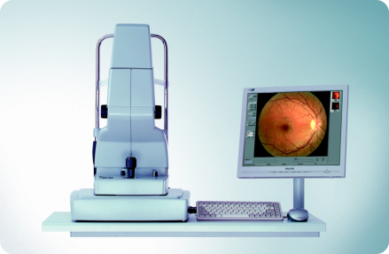
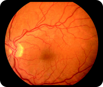
Digital Retinal photography adds a number of diagnostic benefits to traditional ophthalmoscopy. After
reviewing and testing a number of systems we decided to settle on the most advanced system available
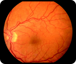
from Zeiss. Our new digital retinal photography system gives your optometrist a high definition view of a
very much larger area of the retina and optic nerve head. This has a number of added benefits in that the
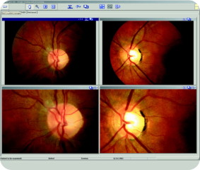
image can be recalled and compared with future findings as a means of detecting slow-changing conditions much earlier. The image can be ‘zoomed’ for even
closer inspection, and manipulated in various ways to show details that cannot be seen by using any other method. It also allows the optometrist to study the
retina as a whole rather than in several smaller views.
Why is it recommended?
Since many conditions that affect the eye can only be detected by examining the retina, it
makes sense to use the best method of inspection and comparison. Our new equipment
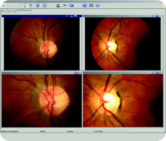
records in accurate detail the appearance of the internal structures of the eyes. Because
changes that happen in conditions like glaucoma or diabetic retinopathy occur very subtly,
the abnormalities are difficult to detect without having something to compare the
appearance against. This ability to store and recall such high quality images is a major step
forward. Although there are obviously differences, the closest analogy to this advancement
would be the use of x-rays in dentistry, or MRI scanning in the modern hospital. Like these
other diagnostic procedures, the real work is done after the image is captured. All images
are studied, analysed and interpreted by a qualified optometrist.
What is involved?
The process of capturing retinal images usually only takes a few minutes. You will sit at the equipment and
be asked to look at a target. When the instrument is aligned there is a brief flash to illuminate the inside of
your eye. The procedure is then repeated for the other eye. Sometimes more than one image may be
required for each eye.
A few patients (those with small pupils and certain types of cataract) may need to have drops to dilate the
pupils before the pictures are taken. You will not be able to drive home if you have had the drops inserted.
How often should it be carried out, and how much does this cost?
Although recognised as one of the most effective diagnostic procedures, digital retinal photography is not
funded by the National Health Service. Please get in touch to ask us the current fee for this additional
procedure. Although it is entirely optional, we advise that at least an initial baseline set of photographs is
taken, with subsequent photographs perhaps every few years unless there are clinical reasons for greater frequency.
As most people realise,
the eye examination
should be a part of every
persons’ regular health
checks. Apart from sight
correction, regular
examinations allow our
optometrists to inspect
your eyes for numerous
conditions that affect general health as well as those that can
affect your vision. The inspection of the interior of the eye is
carried out during a regular eye examination with a hand-held
instrument called an ophthalmoscope. It provides a highly
magnified view of small areas of the retina. Abnormalities and
small changes over time can be noted and compared with
previous notes.
What is Digital Retinal Photography?
Digital Retinal photography adds a number of diagnostic benefits
to traditional ophthalmoscopy. After reviewing and testing a
number of systems we decided to settle on the most advanced
system available from Zeiss. Our new digital retinal photography
system gives your optometrist a high definition view of a very
much larger area of the retina and optic nerve head. This has a
number of added benefits in that the image can be recalled and
compared with future findings as a means of detecting slow-
changing conditions much earlier. The image can be ‘zoomed’ for
even closer inspection, and manipulated in various ways to show
details that cannot be seen by using any other method. It also
allows the optometrist to study the retina as a whole rather than
in several smaller views.
Why is it recommended?
Since many conditions that affect the eye can only be detected by
examining the retina, it makes sense to use the best method of
inspection and comparison. Our new equipment records in
accurate detail the appearance of the internal structures of the
eyes. Because changes that happen in conditions like glaucoma
or diabetic retinopathy occur very subtly, the abnormalities are
difficult to detect without having something to compare the
appearance against. This ability to store and recall such high
quality images is a major step forward. Although there are
obviously differences, the closest analogy to this advancement
would be the use of x-rays in dentistry, or MRI scanning in the
modern hospital. Like these other diagnostic procedures, the real
work is done after the image is captured. All images are studied,
analysed and interpreted by a qualified optometrist.
What is involved?
The process of capturing retinal images usually only takes a few
minutes. You will sit at the equipment and be asked to look at a
target. When the instrument is aligned there is a brief flash to
illuminate the inside
of your eye. The
procedure is then
repeated for the
other eye.
Sometimes more
than one image may
be required for each
eye.
A few patients (those with small pupils and certain types of
cataract) may need to have drops to dilate the pupils before the
pictures are taken. You will not be able to drive home if you have
had the drops inserted.
How often should it be carried out, and how much does this
cost?
Although recognised as one of the most effective diagnostic
procedures, digital retinal photography is not funded by the
National Health Service. Please get in touch to ask us the current
fee for this additional procedure. Although it is entirely optional,
we advise that at least an initial baseline set of photographs is
taken, with subsequent photographs perhaps every few years
unless there are clinical reasons for greater frequency.