During your eye examination, you may have been offered an OCT scan of your eyes and
wondered what this new scan was all about. OCT (optical coherence tomography) is a new,
completely painless and advanced imaging system suitable for people of all ages. It checks for
potentially serious conditions such as glaucoma, diabetes, macular degeneration, vitreous
detachments and more.
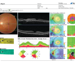
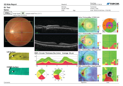
Similar to ultrasound, OCT uses light rather than sound waves to image the different layers that
make up the structures of your eye. The OCT machine captures both a photograph and a cross-
sectional scan of the eye at the same time, enabling your optometrist to see deep into the
tissues and structures of the eye.
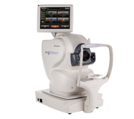
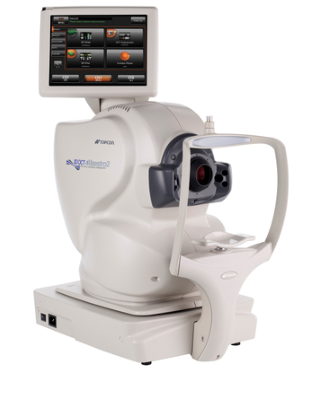
Using our new state-of-the-art 3D OCT system, we will take both a digital photograph and a
three dimensional cross sectional scan of the back of your eye in one sitting. This allows both
instant and early diagnosis of a number of common ocular conditions. The scan is non-invasive, painless, simple and quick. What’s more, the software can
automatically detect even the most subtle changes to the retina with every eye test you have. This gives you an invaluable ongoing record of the health and
condition of your eyes.
OCT scans can detect a large number of conditions that may affect many different structures within the eye.
Common conditions that can be identified and monitored through regular OCT screening include:
AGE-RELATED MACULAR DEGENERATION
Age-related macular degeneration (AMD) is the leading cause of blindness in the UK. It causes gradual
deterioration of the macula (the central portion of your retina which enables detailed vision). There are two
types of AMD; dry and wet. Wet AMD can caus rapid reduction in vision and must be treated in hospital
rapidly. OCT can help to identify the earliest signs of AMD, determine whether it is the dry or wet form and
help monitor its progress over time.
DIABETES
Over 4 million people are now diagnosed with diabetes in the UK, with experts claiming that over half a
million people are currently suffering from undiagnosed type 2 diabetes. Diabetic Retinopathy is one of the
leading causes of blindness in people of working age within the UK. OCT examination helps enable early
detection of diabetic retinopathy, allowing early referral and management which can greatly improve the
success rate of treatment.
GLAUCOMA
Glaucoma is a condition which causes damage to the optic nerve – the part of the eye which connects to the
brain – and causes gradual loss in peripheral vision. Recent statistics suggest that some form of glaucoma
affects around one in 50 people over the age of 40, rising to almost one in 10 in people over 75 years.
Because the early stages of chronic glaucoma do not cause symptoms, regular eye examinations are essential
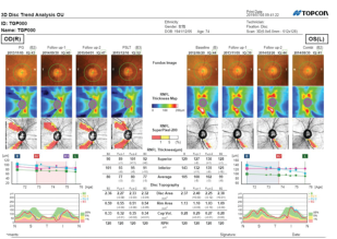
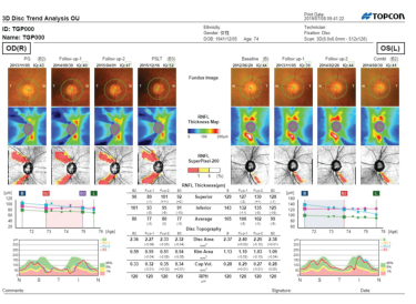
to pick up glaucoma at its earliest stage so that ongoing damage can be prevented. OCT examination can
measure numerous features at the back of the eye and facilitate early diagnosis of glaucoma – often several
years before it can be detected by other means. Furthermore, it can enable close monitoring of your eye
health year-on-year, allowing identification and surveillance of glaucomatous changes over time.
VITREOUS DETACHMENTS
Vitreomacular traction can be easily diagnosed through OCT providing invaluable information about the current relationship between the vitreous and the
retinal surface of the eye. As people get older the vitreous jelly that takes up the space in our eyeball can change. It becomes less firm and can move away from
the back of the eye towards the centre, in some cases parts do not detach and cause ‘pulling’ of the
retinal surface. The danger of a vitreous detachment is that there is no pain and your eyesight will
seem unchanged but the back of your eye may be being damaged.
MACULAR HOLES
As the name suggests, a macular hole is a small hole in the macula – the part of the retina which is
responsible for our sharp, detailed central vision. This is the vision we use when looking directly at
things, when reading, sewing or using a computer for example. Macular holes usually form during a
complicated vitreous detachment, when the vitreous pulls away from the back of the eye, causing a
hole to form. OCT is able to detect these movements enabling rapid referral for management by an
ophthalmologist in hospital.
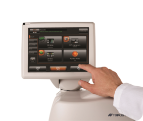
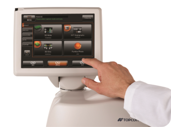
Never before has it been easier to locate and identify eye conditions that affect the layers below the surface. Using our 3D OCT linked to specialised computer
equipment, your optometrist can review the images on a screen and pinpoint areas of concern. Using this equipment, your optometrist can more confidently
diagnose, manage or refer you to a hospital specialist for further treatment, should this be required. For the majority of patients, OCT provides significant
reassurance that the deeper layers of their eye are healthy.
During your eye
examination, you may
have been offered an
OCT scan of your eyes
and wondered what this
new scan was all about.
OCT (optical coherence
tomography) is a new,
completely painless and
advanced imaging system suitable for people of all ages. It checks
for potentially serious conditions such as glaucoma, diabetes,
macular degeneration, vitreous detachments and more.
Similar to ultrasound, OCT uses light rather than sound waves to
image the different layers that make up the structures of your
eye. The OCT machine captures both a photograph and a cross-
sectional scan of the eye at the same time, enabling your
optometrist to see deep into the tissues and structures of the eye.
Using our new state-of-the-art 3D OCT system, we will take both a
digital photograph and a three dimensional cross sectional scan
of the back of your eye in one sitting. This allows both instant and
early diagnosis of a number of common ocular conditions. The
scan is non-invasive, painless, simple and quick. What’s more, the
software can automatically detect even the most subtle changes
to the retina with every eye test you have. This gives you an
invaluable ongoing record of the health and condition of your
eyes.
OCT scans can detect a large number of conditions that may
affect many different structures within the eye.
Common conditions that can be identified and monitored
through regular OCT screening include:
AGE-RELATED MACULAR
DEGENERATION
Age-related macular
degeneration (AMD) is
the leading cause of
blindness in the UK. It
causes gradual
deterioration of the
macula (the central
portion of your retina which enables detailed vision). There are
two types of AMD; dry and wet. Wet AMD can caus rapid
reduction in vision and must be treated in hospital rapidly. OCT
can help to identify the earliest signs of AMD, determine whether
it is the dry or wet form and help monitor its progress over time.
DIABETES
Over 4 million people are now diagnosed with diabetes in the UK,
with experts claiming that over half a million people are currently
suffering from undiagnosed type 2 diabetes. Diabetic Retinopathy
is one of the leading causes of blindness in people of working age
within the UK. OCT examination helps enable early detection of
diabetic retinopathy, allowing early referral and management
which can greatly improve the success rate of treatment.
GLAUCOMA
Glaucoma is a condition which causes damage to the optic nerve
– the part of the eye
which connects to the
brain – and causes
gradual loss in
peripheral vision.
Recent statistics
suggest that some
form of glaucoma
affects around one in
50 people over the
age of 40, rising to almost one in 10 in people over 75 years.
Because the early stages of chronic glaucoma do not cause
symptoms, regular eye examinations are essential to pick up
glaucoma at its earliest stage so that ongoing damage can be
prevented. OCT examination can measure numerous features at
the back of the eye and facilitate early diagnosis of glaucoma –
often several years before it can be detected by other means.
Furthermore, it can enable close monitoring of your eye health
year-on-year, allowing identification and surveillance of
glaucomatous changes over time.
VITREOUS DETACHMENTS
Vitreomacular traction can be easily diagnosed through OCT
providing invaluable information about the current relationship
between the vitreous and the retinal surface of the eye. As people
get older the vitreous jelly that takes up the space in our eyeball
can change. It becomes less firm and can move away from the
back of the eye towards the centre, in some cases parts do not
detach and cause ‘pulling’ of the retinal surface. The danger of a
vitreous detachment is that there is no pain and your eyesight will
seem unchanged but the back of your eye may be being
damaged.
MACULAR HOLES
As the name suggests, a macular hole is a small hole in the
macula – the part of the retina which is responsible for our sharp,
detailed central
vision. This is the
vision we use
when looking
directly at things,
when reading,
sewing or using a
computer for
example. Macular
holes usually form
during a complicated vitreous detachment, when the vitreous
pulls away from the back of the eye, causing a hole to form. OCT
is able to detect these movements enabling rapid referral for
management by an ophthalmologist in hospital.
Never before has it been easier to locate and identify eye
conditions that affect the layers below the surface. Using our 3D
OCT linked to specialised computer equipment, your optometrist
can review the images on a screen and pinpoint areas of concern.
Using this equipment, your optometrist can more confidently
diagnose, manage or refer you to a hospital specialist for further
treatment, should this be required. For the majority of patients,
OCT provides significant reassurance that the deeper layers of
their eye are healthy.